Leg Bone Diagram Labeled - Broken Ankle Types Of Fractures Diagnosis Treatments - The patella (kneecap) is the sesamoid bone in front of the knee.. A labeled diagram of the knee with an insight into its working. Joints and bones of the skeleton pelvis, thigh, hip, knee, leg, ankle and foot were also labeled. In humans the neck of the femur connects the shaft and head at a 125 degree angle, which is efficient for walking. Terms in this set (7) femur. This page is about leg bones diagram,contains aluminium plant safety:
/ this lengthy bone connects with the knee at. Start studying leg bone anatomy. The knee joint, you need a perfectly labeled diagram of the knee. Leg bone anatomy diagram diagram of human leg human anatomy. Tibia and fibula mnemonic to help you learn them, as well as detailed description of the major anatomical structures on each leg bone.
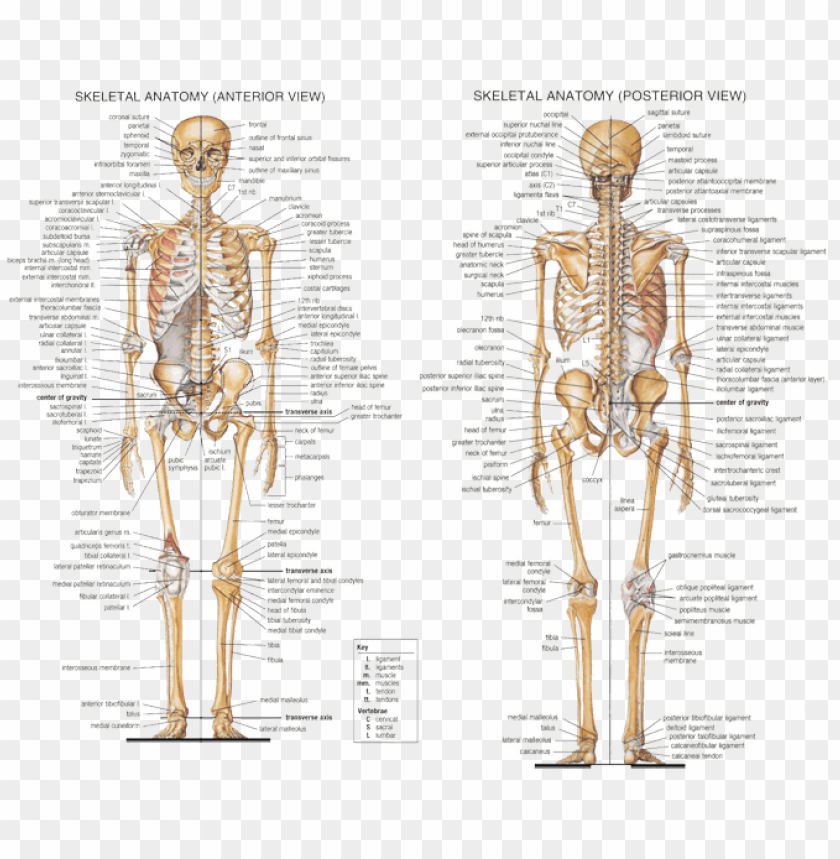
Anatomy Axial Skeleton Human Skeleton All Bones Labeled Png Image With Transparent Background Toppng from toppng.com The second largest bone in physique is the tibia, additionally known as the shinbone. Master leg and knee anatomy using our topic page. Femur, upper bone of the leg or hind leg. However, the definition in human anatomy refers only to the section of the lower limb. Health diagram bone skeleton leg knee science anchor chart human human body. Review date 7/8/2020 updated by: Bone diagram forehead (frontal bone) nose bones (nasals) cheek bone (zygoma) upper jaw (maxilla) lower jaw (mandible) breast bone (sternum) upper arm bone (humerus) lower arm bone (ulna) thigh bone (femur) collar bone (clavicle) toe bones (phalanges) ankle bones (tarsals) kneecap (patella) shin bone Formed by the left and right hip bones, the pelvic girdle connects the lower limb (leg) bones to the axial skeleton.
At the end this module please note that anatomical conventional radiology could be useful for usual practice, which is why you may find some labeled digital radiographs.
/ this lengthy bone connects with the knee at. Beside that, we also come with more related ideas as follows free printable human anatomy coloring pages, lower leg muscle diagram blank and lower limb bones unlabeled. The thigh bone, or femur, is the large upper leg bone that connects the lower leg bones (knee joint) to the pelvic bone (hip joint). The foot bones shown in this diagram are the talus, navicular, cuneiform, cuboid, metatarsals and calcaneus. Health diagram bone skeleton leg knee science anchor chart human human body. Each leg is made up of four bones. It's the area that runs from the hip to the knee in each leg. This image is an edited version of this image that was created by user:ladyofhats (mariana ruiz villarreal). A heavy, long bone that forms the leg above the knee. The foot bones shown in this diagram one of the beloved. The tibia (also called the shinbone) is located near the midline of the leg. The upper leg is often called the thigh. Posted on april 18, 2019april 18, 2019.
However, the definition in human anatomy refers only to the section of the lower limb. Learn vocabulary, terms, and more with flashcards, games, and other study tools. The foot bones shown in this diagram one of the beloved. {label gallery} get some ideas to make labels for bottles, jars, packages, products, boxes or classroom activities for free. Beside that, we also come with more related ideas as follows free printable human anatomy coloring pages, lower leg muscle diagram blank and lower limb bones unlabeled.
Human Skeleton Coloring Page Medical Art Library from www.medicalartlibrary.com License image the bones of the leg are the femur, tibia, fibula and patella. This image is an edited version of this image that was created by user:ladyofhats (mariana ruiz villarreal). Hip and leg bone diagram : The femur is the largest bone in the body and the only bone of the thigh. The bones together make up the hip. Our goal is that these leg anatomy worksheets pictures gallery can be a direction for you, bring you more references and also make you have a great day. The head of the femur forms the ball and socket hip joint with the acetabulum of the hip bone. The bones of the leg are the femur, tibia, fibula and patella.
Our goal is that these leg anatomy worksheets pictures gallery can be a direction for you, bring you more references and also make you have a great day.
Its lower end helps create the knee joint. The bones of the leg are the femur, tibia, fibula and patella. To understand one of the most complex joints of our body i.e. The knee joint, you need a perfectly labeled diagram of the knee. Hip and leg bone diagram : Tibia and fibula bone anatomy with diagram picture and free quiz. The hip itself is a ball and socket joint, much like the shoulder.the structures necessary to create this joint are the socket, the joint capsule, muscle, ligaments, and the neck. However, the definition in human anatomy refers only to the section of the lower limb. Our goal is that these leg anatomy worksheets pictures gallery can be a direction for you, bring you more references and also make you have a great day. Review date 7/8/2020 updated by: The knee joint is the largest joint in the body and is primarily a hinge joint, although some sliding and rotation occur. / this lengthy bone connects with the knee at. A labeled diagram of the knee with an insight into its working.
The hip joint gives the leg an incredible range of motion while still providing support to the body's weight. The knee joint is the largest joint in the body and is primarily a hinge joint, although some sliding and rotation occur. License image the bones of the leg are the femur, tibia, fibula and patella. The foot bones shown in this diagram are the talus, navicular, cuneiform, cuboid, metatarsals and calcaneus. Review date 7/8/2020 updated by:
Lateral View Of Male Pelvis Hip And Leg Bones Labeled On A White Background Stocktrek Images from www.stocktrekimages.com The bones of the leg are the femur, tibia, fibula and patella. Long bones are found in the arms (humerus, ulna, radius) and legs (femur, tibia, fibula), as well as in. Master leg and knee anatomy using our topic page. Review date 7/8/2020 updated by: Knee, leg, and foot (overview) how many times have a layman's language and anatomy ever matched? Beside that, we also come with more related ideas as follows free printable human anatomy coloring pages, lower leg muscle diagram blank and lower limb bones unlabeled. The knee joint, you need a perfectly labeled diagram of the knee. Each leg is made up of four bones.
Labeled human leg bones created for use in leg bone.
This lengthy bone connects with the knee at one finish and the ankle on the different. Formed by the left and right hip bones, the pelvic girdle connects the lower limb (leg) bones to the axial skeleton. Learn with flashcards, games, and more — for free. The femur, or thighbone, is the longest and largest bone in the human body. Terms in this set (7) femur. To understand one of the most complex joints of our body i.e. The bones of the hip include the femur, the ilium, the ischium, and the pubis. Learn vocabulary, terms, and more with flashcards, games, and other study tools. The head of the femur forms the ball and socket hip joint with the acetabulum of the hip bone. Together, these muscles straighten your knee, stabilize your knee joint, assist in flexing your hip (drawing your knee towards your chest), and help absorb force when you land after jumping or leaping. The anatomy of the leg and foot bones. The anatomy of the leg and foot bones. The hip joint gives the leg an incredible range of motion while still providing support to the body's weight.
The knee joint, you need a perfectly labeled diagram of the knee leg bone diagram. The head of the femur forms the ball and socket hip joint with the acetabulum of the hip bone.